
High Screen Time: New Macular Carotenoids Supplement Study Supports Eye Health

By Thomas A. Bowman, PhD
The average American spends over 10 hours a day looking at smart phones, tablets or computer screens. All of that time looking at a screen can have a cumulative effect on your eyes. A new study, published recently in the journal Foods has found that supplementation with macular carotenoids improved vision, sleep quality and other physical symptoms in young healthy adults who had a high exposure to "screen time" (those using smart phones, tablets or computers more than 6 hours daily).1*
Visual measurements in the study included significantly improved contrast sensitivity, decreased glare and improved photostress recovery.* Along with these visual improvements and improved sleep quality, subjects also had significantly fewer headaches, and less eyestrain and eye fatigue.* As found in many previous studies, macular carotenoid supplementation significantly improved serum levels and macular pigment optical density.1
Macular Carotenoids -Lutein, Zeaxanthin and Mesozeaxanthin
|
Macular carotenoids are pigments, derived from the diet. These carotenoids include lutein, zeaxanthin and mesozeaxanthin. |
|
 |
The macula (see Figure 1) is the central focal point of the retina of the eye that requires a very high concentration of these macular carotenoids. Accordingly, the macula is especially important for color vision and high definition central vision. How macular carotenoids protect individuals with high screen time is due to their capability as antioxidants and in blue light filtering (discussed further below). Figure 1: The macula and retina |
What are Carotenoids?
Carotenoids produce the vibrant colors found in many vegetables. The name carotenoid brings to mind the similarly sounding vegetable, the carrot. Carrots indeed contain carotenoids, primarily beta-carotene and alpha- carotene, which give the carrot its orange color. Other carotenoids in foods include lycopene and beta- cryptoxanthins. Our mothers told us as children to eat our carrots to help our vision. However, macular carotenoids (not found in carrots) are particularly important for vision.
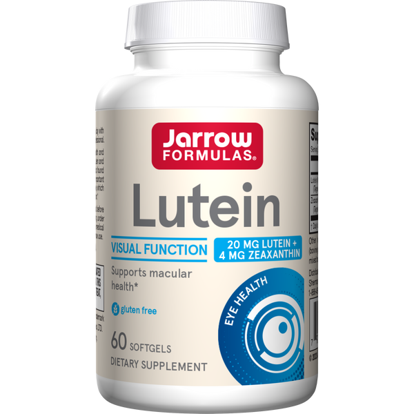
Lutein, found in leafy green vegetables such as kale and spinach, is the most abundant macular carotenoid in the body. Zeaxanthin, found in corn and eggs, is also an important macular carotenoid. Only the diet provides lutein and zeaxanthin. The body is unable to make these two macular carotenoids. In contrast, the macula makes mesozeaxanthin from lutein.2 The density of macular carotenoids in the maculae and in blood varies in individuals depending on dietary intake.3 Consequently, many individuals have had no detected macular carotenoids.3
Not restricted to the eyes, macular carotenoids are in many places throughout the body, but especially in fatty tissues like fat and the brain. A prior article discussed the role of lutein in infant brains, based on a study of significant lutein accumulation in the brains of infant rhesus macaques after taking lutein-supplemented infant formula.4 In another article, we explained a recent study5 on the important role of macular carotenoids in brain cognitive function and psychological stress.* Unfortunately, being overweight decreases the amount of macular carotenoids that make it to the eyes, since fat tissue in the rest of the body also takes up the macular carotenoids.6
Structure of Macular Carotenoids for Blue Light Filtering
Macular carotenoids filter blue light, because of their physical property as dark yellow pigments. Blue light is especially important in the central macula of the eye due to the retina allowing increased blue light to enter this area. The increased blue light in the macula is due to the absence of several neural cell layers. Blue cones are absent in the central part of the macula. Thus, the macula needs a substantial density of macular carotenoids here to filter any blue light to protect the photoreceptor membranes.
| The filtration of blue light depends on the orientation of macular carotenoid molecules. Their molecular structure allows macular carotenoids to span the membrane. Macular carotenoids are straight molecules with a rigid middle bar region that is hydrophobic and hydroxyls at each end that are more hydrophilic (see Figure 2). This is unlike cholesterol, which extends only into half of the membrane, or other carotenoids that are fully hydrophobic. |
Figure 2: The structure of Macular Carotenoids (modified from Widomska 7) |

 |
The ability of macular carotenoids to span the membrane makes them more stable and therefore, longer lasting.3 Spanning the membrane also orients these pigments perpendicular to the direction of blue light entering the photoreceptors (see Figure 3). This perpendicular orientation makes them perfect for filtering blue light. Figure 3: Light orientation on photoreceptors |
Function of Macular Carotenoids for Antioxidant Blue Light Decontamination
As previously described, the macula is especially concentrated with high definition neural photoreceptor cone cells, responsible for highly focused and brightly colored central vision. The increased blue light to enter this area makes it most susceptible to photo-oxidation.* Photoreceptors have blue light absorbing molecules that allow absorption of blue light, which produces chain reactions of free radicals and reactive oxygen in these photoreceptors.*
The membranes of the photoreceptors are particularly vulnerable to these high-energy reactions. Macular carotenoids concentrated in this area also decontaminate the membranes by scavenging free radicals, neutralizing singlet oxygen, and vibrating to release the energy. Membranes consist mainly of phospholipids that free radicals can convert into lipid peroxides. Several studies have shown the protective effects of macular carotenoids in preventing formation of these lipid peroxides from blue light (reviewed by Bernstein).8*
References:
- Stringham JM, Stringham NT, O’Brien KJ. Macular Carotenoid Supplementation Improves Visual Performance, Sleep Quality, and Adverse Physical Symptoms in Those with High Screen Time Exposure. Foods. 2017;6 (7):47.
- BONE RA, LANDRUM JT, FRIEDES LM, et al. Distribution of lutein and zeaxanthin stereoisomers in the human retina. Experimental eye research. 1997;64(2):211-218.
- Curran-Celentano J, Hammond BR, Ciulla TA, Cooper DA, Pratt LM, Danis RB. Relation between dietary intake, serum concentrations, and retinal concentrations of lutein and zeaxanthin in adults in a Midwest population. The American journal of clinical nutrition. 2001;74(6):796-802.
- Jeon S, Neuringer M, Johnson EE, et al. Effect of carotenoid supplemented formula on carotenoid bioaccumulation in tissues of infant rhesus macaques: A pilot study focused on lutein. Nutrients. 2017;9 (1):51.
- Stringham NT, Holmes PV, Stringham JM. Supplementation with macular carotenoids reduces psychological stress, serum cortisol, and sub-optimal symptoms of physical and emotional health in young adults. Nutritional Neuroscience. 2017:1-11.
- Nolan J, O’Donovan O, Kavanagh H, et al. Macular pigment and percentage of body fat. Investigative ophthalmology & visual science. 2004;45(11):3940-3950.
- Widomska J, Zareba M, Subczynski WK. Can xanthophyll-membrane interactions explain their selective presence in the retina and brain? Foods. 2016;5(1):7.
- Bernstein PS, Khachik F, Carvalho LS, Muir GJ, Zhao D-Y, Katz NB. Identification and quantitation of carotenoids and their metabolites in the tissues of the human eye. Experimental eye research. 2001;72 (3):215-223.


